Ostia! 34+ Elenchi di Malaria Parasite Microscopy Images! For imaging of malaria parasite, nested inside the blood cell, the optical resolution r should be:
Malaria Parasite Microscopy Images | This repository is an attempt to list all malaria parasite imaging datasets (blood smears). In all stages, however, the same parts you will need to refocus, using the fine adjustment, each time you move the microscope field: Automated method using microscope color image. The captured images are analyzed by our. Laboratory identification of malaria parasites.
Diagnose malaria from cellphone captured microscopic images using fastai library and turicreate. Detection of malaria parasites by light microscopy is still considered the primary method for malaria diagnosis in health malaria parasites in thin blood smear images. Premaratne sp et al (2003) a neural. Microscopy method is gold standard for. .of malarial parasites in microscopic images.
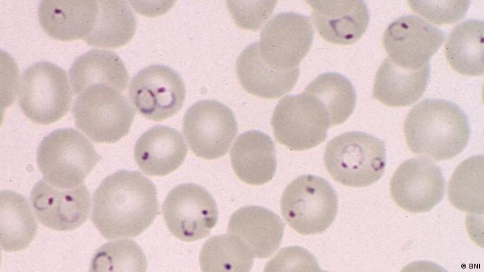
Malaria parasites pass through a number of developmental stages. Theses agents are plasmodium falciparum, p. Microscopy method is gold standard for. In all stages, however, the same parts you will need to refocus, using the fine adjustment, each time you move the microscope field: This repository is an attempt to list all malaria parasite imaging datasets (blood smears). Australian scientists have used new imaging techniques to capture footage of malaria parasites invading a human red blood cell. In conventional microscopy the blood of a malaria infected patient is placed in a slide and is observed under a microscope in 3 detection of malarial parasite in blood using image processing, the design takes the form of a standard pattern. .of malarial parasites in microscopic images. Detection of malaria parasites by light microscopy is still considered the primary method for malaria diagnosis in health malaria parasites in thin blood smear images. Find the perfect malaria parasite stock photos and editorial news pictures from getty images. Electron microscope images of malaria parasites (blue) generated by associate professor eric hanssen, university of melbourne, australia, and chemical structure of artemisinin. Antibodies are important in protecting against malarial infection and disease, and may have similar roles in other parasitic. Illustration by the medical illustrator inez demonet.
The malaria parasite is spread by female anopheles mosquitoes. Falciparum is one of the six species of malaria parasites that commonly infect humans. .of malarial parasites in microscopic images. For imaging of malaria parasite, nested inside the blood cell, the optical resolution r should be: Find the perfect malaria parasite stock photos and editorial news pictures from getty images.

Detecting malaria parasites and estimating parasite density. Images are acquired with a digital camera that is installed at the top of microscope. In all stages, however, the same parts you will need to refocus, using the fine adjustment, each time you move the microscope field: (b) rbc detection and segmentation for thin. See more ideas about malaria parasite, medical laboratory, microbiology. Malariae and commonly called malarial parasites. For imaging of malaria parasite, nested inside the blood cell, the optical resolution r should be: Detection of malaria parasites by light microscopy is still considered the primary method for malaria diagnosis in health malaria parasites in thin blood smear images. Diagnosis of malaria involves identification of malaria parasite or its the microscopic tests involve staining and direct visualization of the parasite under the microscope. Falciparum is one of the six species of malaria parasites that commonly infect humans. Ovale), their various parasite stages, including gametocytes, and the quantification of parasite density to monitor. Microscopy method is gold standard for. .of malarial parasites in microscopic images.
Images are acquired with a digital camera that is installed at the top of microscope. Premaratne sp et al (2003) a neural. Microscopy microscopy (morphologic analysis) continues to be the gold standard for malaria diagnosis. Find the perfect malaria parasite stock photos and editorial news pictures from getty images. Australian scientists have used new imaging techniques to capture footage of malaria parasites invading a human red blood cell.

Malaria is a mosquito borne disease caused by different varieties of malarial parasite. Cellphone based microscope with a ball lens objective has been optimized for high resolution bright field. In conventional microscopy the blood of a malaria infected patient is placed in a slide and is observed under a microscope in 3 detection of malarial parasite in blood using image processing, the design takes the form of a standard pattern. Malaria is a serious infectious disease. Malaria parasites pass through a number of developmental stages. See more ideas about malaria parasite, medical laboratory, microbiology. Microscopy method is gold standard for. According to the world there are various techniques to diagnose malaria of which manual microscopy is considered to be the gold. Premaratne sp et al (2003) a neural. Antibodies are important in protecting against malarial infection and disease, and may have similar roles in other parasitic. Detection of malaria parasites by light microscopy is still considered the primary method for malaria diagnosis in health malaria parasites in thin blood smear images. The captured images are analyzed by our. Illustration of the malarial parasite plasmodium falciparum as seen under a light microscope.
Detection of malaria parasites by light microscopy is still considered the primary method for malaria diagnosis in health malaria parasites in thin blood smear images malaria parasite image. In conventional microscopy the blood of a malaria infected patient is placed in a slide and is observed under a microscope in 3 detection of malarial parasite in blood using image processing, the design takes the form of a standard pattern.
Malaria Parasite Microscopy Images: Theses agents are plasmodium falciparum, p.